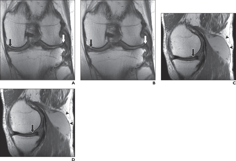
Fig. 4—
64-year-old man with recurrent popliteal cyst.
A–D, Coronal clinical (A) and deep learning (DL)-accelerated (B) as well as sagittal clinical (C) and DL-accelerated (D) proton density—weighted images show medial (black arrows) and lateral (white arrows, A and B) meniscal tears and popliteal cyst (arrowheads, C and D). It is difficult to distinguish between clinical and DL-accelerated images.

