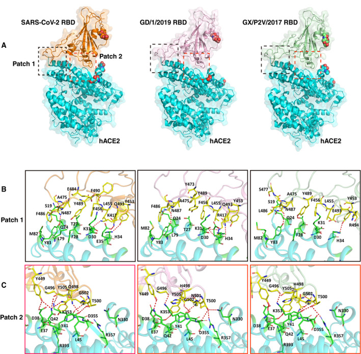
Figure 4. The complex structures of GD/1/2019 RBD and GX/P2V/2017 RBD bound to hACE2.

-
AThe overall complex structures of hACE2 bound to the SARS‐CoV‐2 RBD, GD/1/2019 RBD, and GX/P2V/2017 RBD. The binding between the RBDs and hACE2 is mainly composed of two patches of interactions, and patch 1 and patch 2 are indicated in black and red dashed boxes, respectively. The N‐glycans are shown as spheres. hACE2, SARS‐CoV‐2 RBD, GD/1/2019 RBD, and GX/P2V/2017 RBD are colored in cyan, orange, light pink, and pale green, respectively.
-
B, CDetailed interaction of hACE2 with the SARS‐CoV‐2 RBD, GD/1/2019 RBD, and GX/P2V/2017 RBD in patch 1 and patch 2. Residues involved in the interaction are labeled, and H‐bonds are shown as red dotted lines with a cutoff of 3.5 Å.
