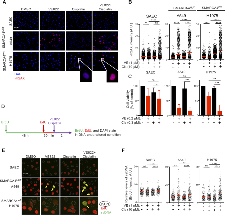
Figure 2.
Cisplatin-mediated DNA replication stress synergistically enhances ATRi-induced RC. (A, B) Cells were treated with the indicated drugs for 8 h and immunostained with anti-γH2AX antibody, and their nuclei were counterstained with DAPI. (A) Representative image. Scale bar: 20 μm. (B) Quantification of the γH2AX intensities of 2000 cells treated with ATRi and cisplatin. Red bars represent mean intensities. Representative results of two independent reproducible experiments are shown. (C) Cells were treated with ATRi and/or cisplatin at the indicated concentrations for 6 days and cell viability was measured by the PrestoBlue assay. Each value represents the mean ± SD of three independent experiments. (D) Schematic representation of the experimental design used to analyze the exposed ssDNA by BrdU staining. Cells were incubated with 10 μM BrdU for 48 h, followed by 10 μM EdU for 30 min and ATRi and/or cisplatin for 2 h. The exposed ssDNA was immunostained with anti-BrdU (green) antibody under native conditions and the S-phase and total nuclei were counterstained with EdU (red) or DAPI (dashed line), respectively. (E) Cells were treated with ATRi and cisplatin as indicated, and the exposed ssDNA was analyzed. Scale bar: 20 μm. (F) Quantification of the ssDNA (BrdU) intensities of 200 S-phase cells (EdU positive) (E). Red bars represent the mean intensity. Representative results of two independent reproducible experiments are shown.

