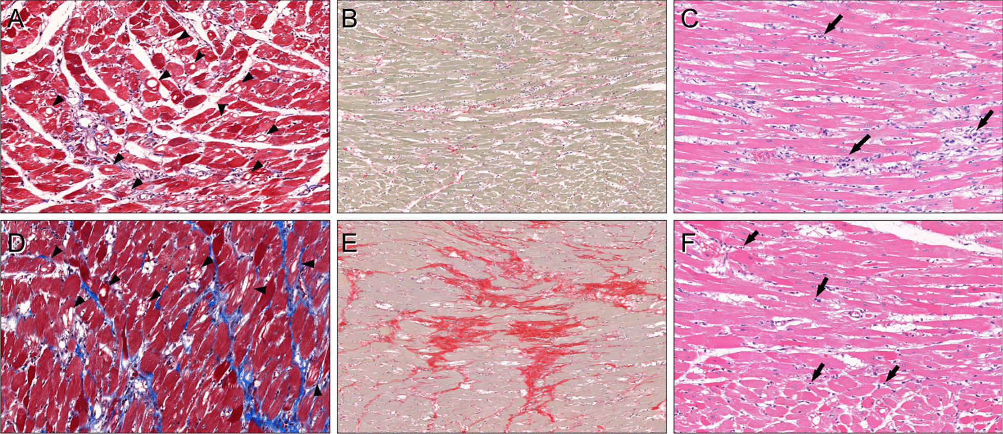
Fig. 5.

CMR data and histopathological findings of doxorubicin-treated rats. a–c For a representative rat at 6 weeks of doxorubicin treatment (group 3) with a left ventricular ejection fraction (LVEF) of 65%, global native T1 of 1401 ms, and ECV of 21.3%, there were marked vacuolar changes (arrowheads) of the myocytes with a mean value of 32.9% (a, Masson’s trichrome stain, × 200). Interstitial fibrosis was 3.0% (b, picrosirius red stain, × 100). There were interstitial edema (median score: 2) and infiltration of lymphohistiocytic aggregates (arrows, median score: 2) around the injured myofibers (c, hematoxylin, and eosin stain, × 200). d–f For a representative rat at 12 weeks of doxorubicin treatment (group 5) with a LVEF of 67%, global native T1 of 1410 ms, and ECV of 28.9%, there were multifocal vacuolar degenerations in the myocytes (arrowheads) with a mean vacuolar change of 38.0% (d, Masson’s trichrome stain, × 200). Interstitial fibrosis was severe, with a mean value of 25.4% (e, picrosirius red stain, × 100). Interstitial edema (median score: 1.75) and scattered lymphocytic infiltration (arrows, median score: 0.25) were also present (f, hematoxylin and eosin stain, × 200)
