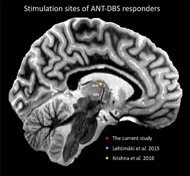
FIGURE 6.

Visual representation of the stimulation sites of ANT-DBS responders in the current study and published studies, overlaid on a sagittal section of a 7T MR image. The sequence used was a T1 white-matter-nulled MPRAGE51 (voxel size: 0.8 × 0.8 × 0.8 mm, TE/TR/TI of 3.3/4.5/617 ms) obtained from a healthy control using a 7T magnet (Siemens, Erlangen, Germany) and a 32-channel head coil (Nova Medical, Wilmington, Massachusetts) at the Maastricht Brain Imaging Centre. Abbreviations: ANT, anterior nucleus of the thalamus; MTT, mammillothalamic tract.
