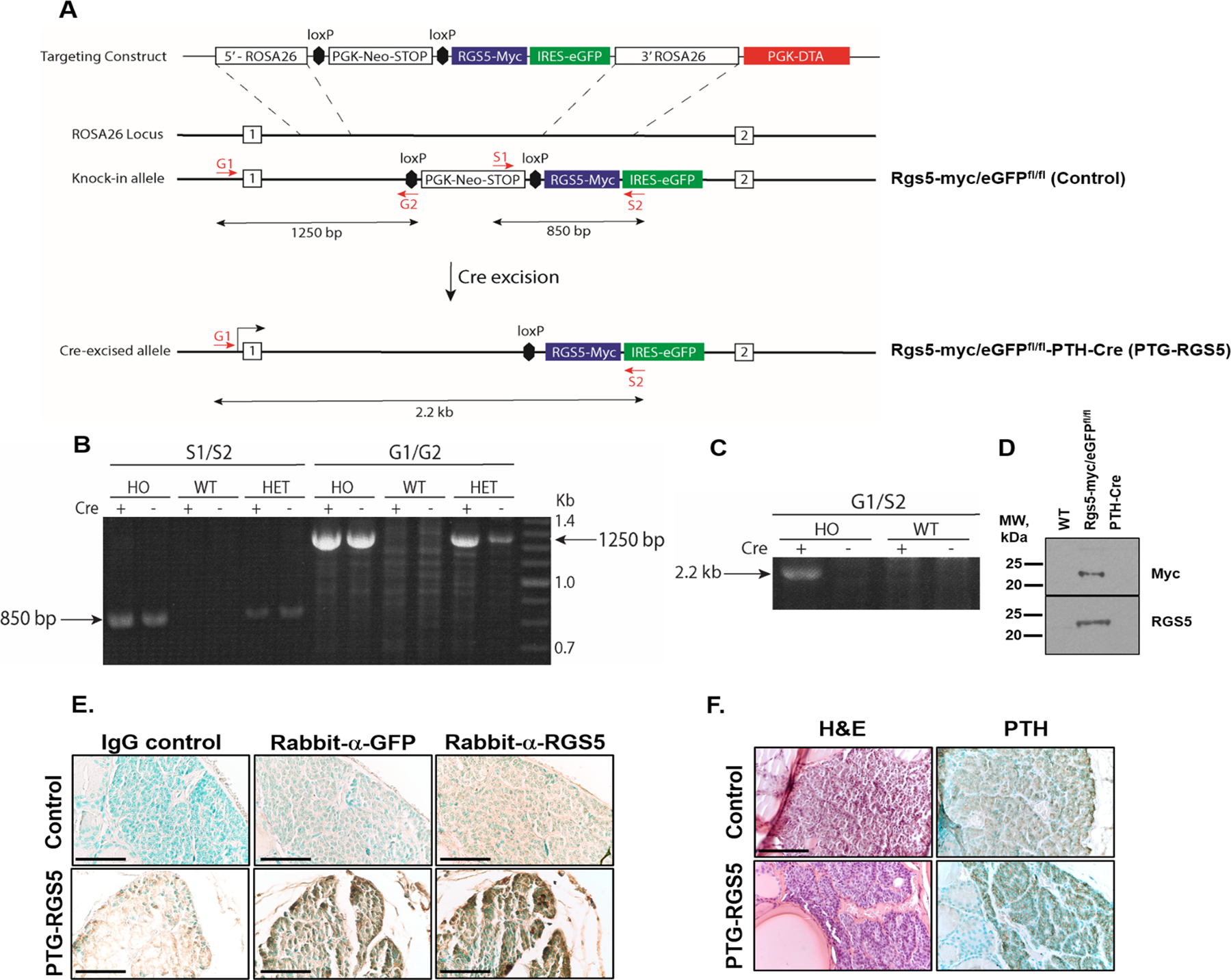
Figure 1.

A. ROSA26 knock-in strategy for creating the parathyroid-specific RGS5 (PTG-RGS5) overexpressing transgenic mouse. B. PCR amplification of the RGS5 transgene from tail snips of homozygous (HO, RGS5-myc/eGFPfl/fl) and heterozygous (HET, RGS5-myc/eGFPfl/+) mice. The S1/S2 primers flank the RGS5 cDNA within the targeting vector and produce an 850 bp product specific to the transgenic mice. The G1/G2 primer combination demonstrates insertion into the ROSA26 locus. PCR from tail snip genomic DNA produces a 1250 bp product indicating correctly oriented transgene insertion in the HO and HET PTG-RGS5 strain. The presence or absence of Cre in mice does not affect the size of either product because the genomic DNA is extracted from tail. C. Cre-dependent recombination at the transgene in PTG-RGS5 mice. Lung fibroblasts from HO PTG-RGS5 or WT mice were transduced in culture with an NLS-Cre lentivirus and then genomic PCR was performed using G1 and S2 primers. In the presence of Cre, the PGK-Neo-STOP cassette is excised, bringing the G1 and S2 sites closer together and allowing amplification of the expected 2.2 kb product. In the absence of NLS-Cre, there is no detectable product. D. Western blot of murine lung fibroblasts from the indicated strains, after infection with the Cre lentivirus. In the RGS5-myc/eGFPfl/fl cells, a band of the correct size is recognized by both an anti-myc monoclonal antibody (9E10) and by an anti-RGS5 polyclonal antibody. E. Mouse tracheal blocks were sectioned and incubated with rabbit IgG control, rabbit anti-GFP or rabbit anti-RGS5 antibody followed by DAB staining (brown) and methylgreen nuclear counterstaining (green). Magnification is 400X and scale bar is 50 μm. F. Mouse tracheal blocks were sectioned and incubated with mouse anti-PTH antibody followed by DAB staining (brown) and methylgreen nuclear counterstaining (green) (right panels) or were stained with hematoxylin/eosin (left panels). Magnification is 400X. Scale bar: 50 μm.
