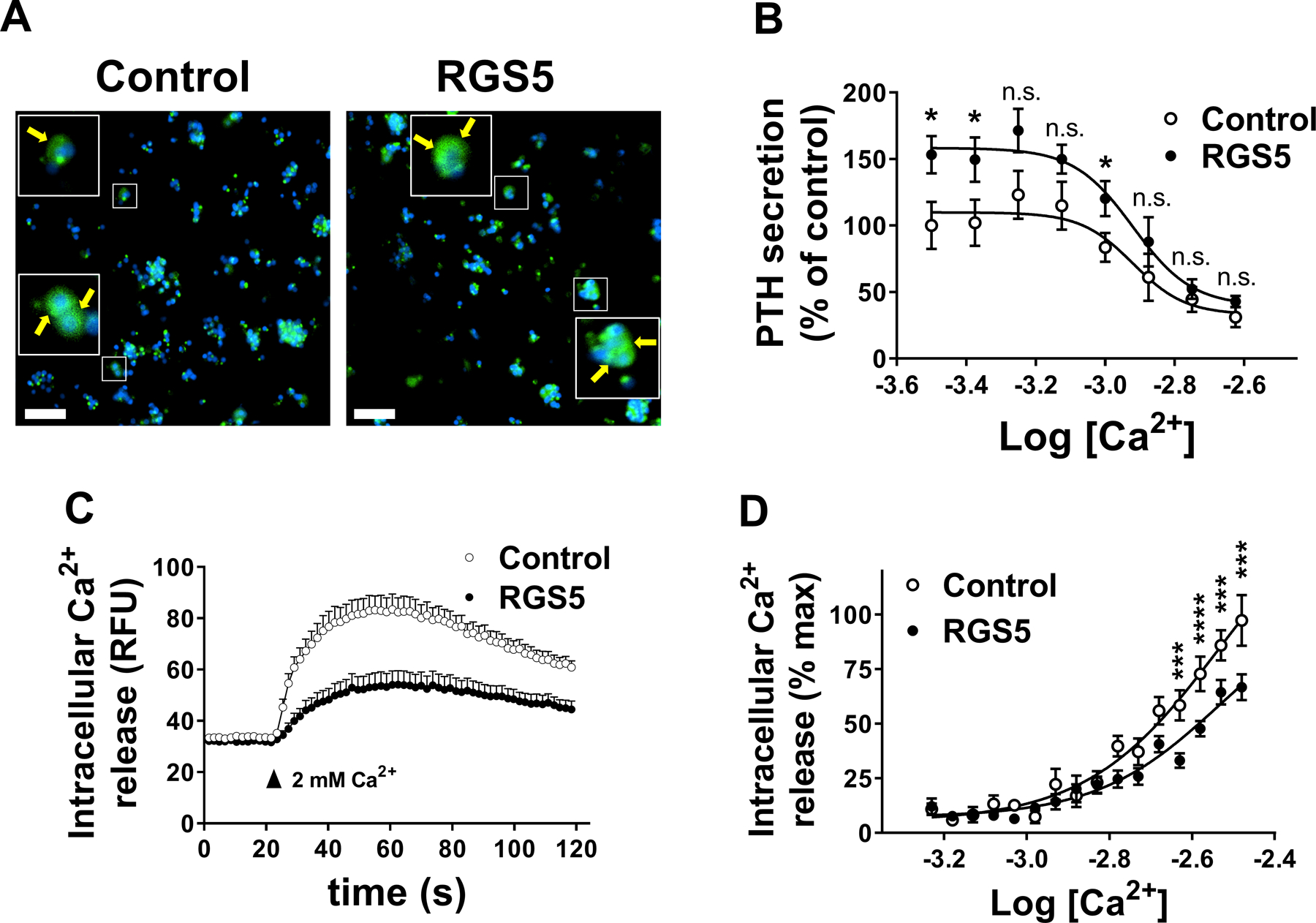
Figure 6.

Effect of overexpression of RGS5 on PTH secretion by normal human parathyroid cells. Dispersed normal cadaveric parathyroid cells from eucalcemic donors were transduced with lentivirus particles expressing GFP reporter along with either LacZ (control) or RGS5 for 24 hrs. A. Cells were fixed and stained with DAPI nuclear dye (blue). GFP (green), co-expressed with transgenes by lentivirus is shown. Inset shows cytoplasmic staining of GFP. Scale bar: 50 μm. B. Cells were stimulated with increasing concentrations of extracellular Ca2+ and PTH secretion was measured in a colorimetric ELISA assay. Data are mean ± SEM from 3 independent experiments performed in triplicate. Nonparametric Mann-Whitney t-test was used for data analysis. *P < 0.05. (n.s. not significant). C. Kinetic of CASR-mediated intracellular Ca2+ release in human normal parathyroid cells was monitored 20 seconds before and 100 seconds after stimulation with 2 mM extracellular Ca2+. D. Concentration response curves of CASR-mediated intracellular calcium release were derived by GraphPad software as described in methods. Data are mean ± SEM from 3 independent experiments conducted in triplicate. Nonparametric Mann-Whitney t-test was used for data analysis. ***P < 0·001, ****P < 0·0001.
