Figure 1.
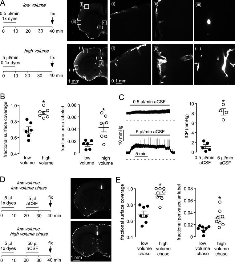
Cisternal injection volume is an important determinant of solute uptake in brain.(A) Left: Low- and high-volume injection protocols in which a fixed amount of fluorescent dextran dissolved in 5 µl (1× dye) or 50 µl (0.1× dye) of aCSF was injected over 10 min. Mice were sacrificed by perfusion fixation 30 min after injection. Right: Fluorescence images showing the distribution of 70-kD fluorescent dextran in brain sections at low resolution (left images), with higher-magnification images in the boxes marked (i)–(iii) (right images). (B) Left: Fraction of brain surface covered by dye (n = 6–7 mice; *, P = 0.0004, t test). Right: Fraction of brain area labeled with dextran (*, P = 0.0063, t test). (C) Left: ICP during cisternal aCSF injections at 0.5 and 5 µl/min. Right: Mean change in ICP during injections (n = 4–5; *, P = 0.0002, t test). (D) Left: Low- and high-volume chase injection protocols, with 5 µl of dye-containing solution injected over 10 min and then chased with 5 or 50 µl aCSF over 10 min. Right: Fluorescence images showing the distribution of 70-kD fluorescent dextran. (E) Left: Fraction of the brain surface covered by dye with low- and high-volume chase protocols (n = 7–8 mice; *, P = 0.0002, t test). Right: Fraction of brain area covered by dye (*, P = 0.0056, t test).

