Figure 6.
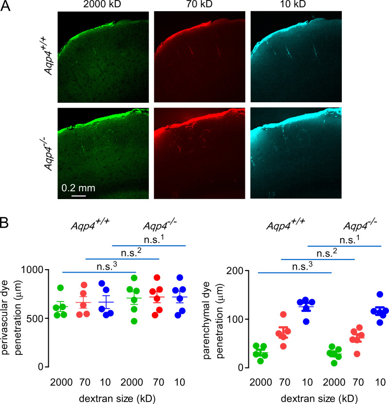
AQP4 deletion does not alter the distribution of fluorescent dextrans in brain following direct application under constant pressure.(A) Images showing distribution of 2,000-, 70-, and 10-kD fluorescent dextrans in brain cortex after direct surface application in wild-type and AQP4 knockout mice. (B) Left: Quantification of dye penetration in the perivascular spaces in mice of each genotype (n.s.1,2, P > 0.999; n.s.3, P = 0.986, repeated-measures two-way ANOVA with Bonferroni posttest; n = 5 AQP4+/+ and 6 AQP4−/− mice). Right: Quantification of dye penetration in the parenchyma in mice of each genotype (n.s.1,3, P > 0.999; n.s.2, P = 0.922, repeated-measures two-way ANOVA with Bonferroni posttest; n = 5 AQP4+/+ and 6 AQP4−/− mice).

