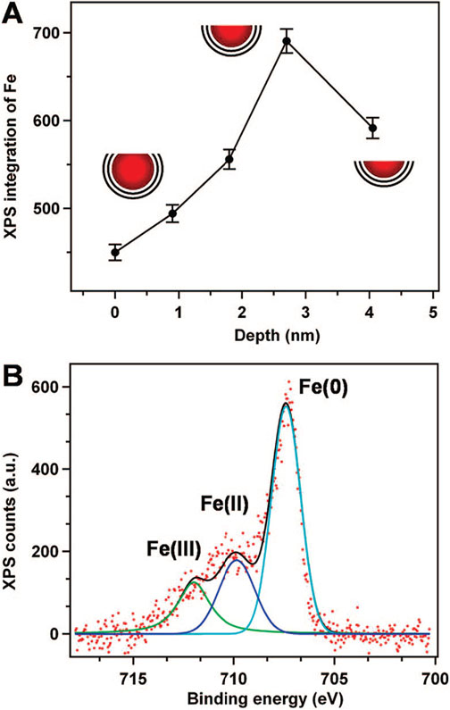
Figure 2.
X-ray photoelectron spectroscopy (XPS) analysis. (A) Iron signal as a function of sputtering time. The sputtering rate was ~0.9 Å/min. (B) XPS of a thin film of NPs on a gold substrate showing Fe(0):Fe(II):Fe(III) ~ 3.4:1.3:1, as inferred from modeling the spectrum with three appropriate oscillators.

