Figure 1.

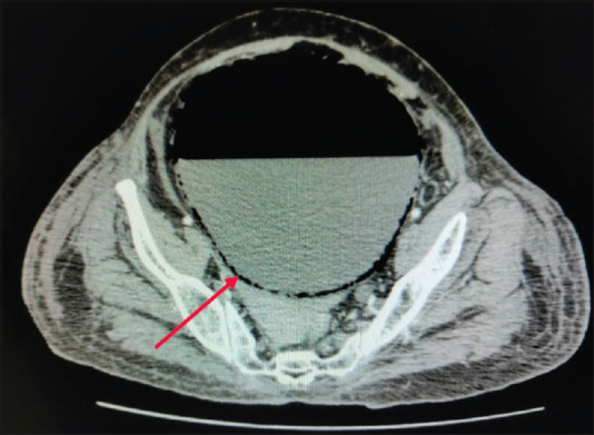
Axial computed tomography scan showing distended bladder with air–fluid level and multiple small foci of air specks in bladder wall (red arrow)

Axial computed tomography scan showing distended bladder with air–fluid level and multiple small foci of air specks in bladder wall (red arrow)