Figure 3.

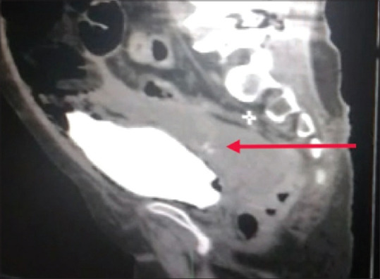
Sagittal computed tomography scan with cystogram showing thin stream of bladder contrast (red arrow) going into intraperitoneal pelvic collection

Sagittal computed tomography scan with cystogram showing thin stream of bladder contrast (red arrow) going into intraperitoneal pelvic collection