
Figure 4.
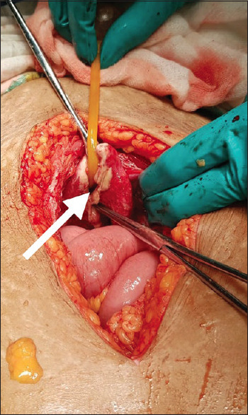
Intraoperative photograph showing bladder perforation near the dome (white arrow) with necrotic sloughed edges with thickened edematous bladder wall and Foley's catheter can be seen coming out through the rent (foot end)
Official websites use .gov
A
.gov website belongs to an official
government organization in the United States.
Secure .gov websites use HTTPS
A lock (
) or https:// means you've safely
connected to the .gov website. Share sensitive
information only on official, secure websites.
Intraoperative photograph showing bladder perforation near the dome (white arrow) with necrotic sloughed edges with thickened edematous bladder wall and Foley's catheter can be seen coming out through the rent (foot end)