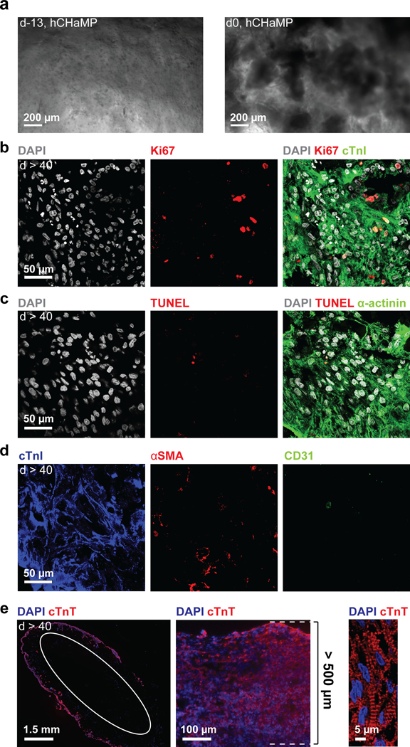
Figure 4. Contiguous muscle formation in hChaMP with ongoing proliferation and limited cell death.

a) Single hiPSCs (d-13) and hiPSC colonies (d0) in hChaMP. b-d) Immunofluorescence staining at six weeks following lactate treatment. The scale is the same for all images. b) Proliferation via Ki67 staining. c) Cell death via TUNEL staining. d) Staining for cardiomyocytes, smooth muscle cells, and endothelial cells in the hChaMP via cTnI, αSMA, and CD31 staining, respectively. e) From left to right: contiguous muscle formation on the majority of the exterior surface of the hChaMP stained for cardiac troponin T (cTnT, white circle indicates inner wall of chamber cross section), a thick region of the hChaMP wall with muscle depth of 500 μm, and high-magnification image of sarcomeric striations within an hChaMP.
