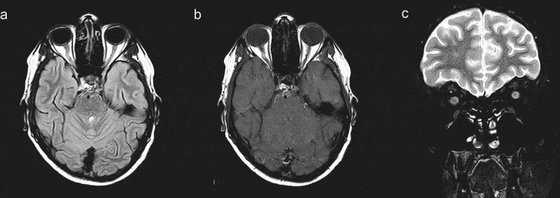
Figure 2.

MRI of the brain and orbits. (a) Axial fast fluid attenuated inversion recovery image showing hyperintensity and increased thickness of the right optic nerve. (b) Gadolinium-enhanced axial T1-weighted image showing enhancement of the right optic nerve head. (c) Coronal T2-weighted image showing an increased diameter of the right optic nerve
