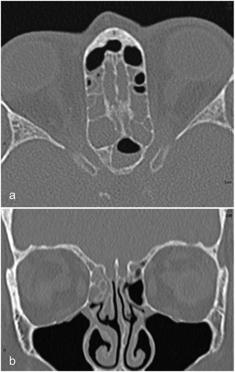
Figure 1.

CT scan of case 1
(a) Transverse reformat from a helical acquisition of the ethmoid and sphenoid sinuses showing unruptured bone. Most ethmoidal cells are obliterated with mucosal thickening and a fluid/air level is seen in the left sphenoid cell; (b) Coronal reformat showing unremarkable mucosal thickening without bone erosion.
