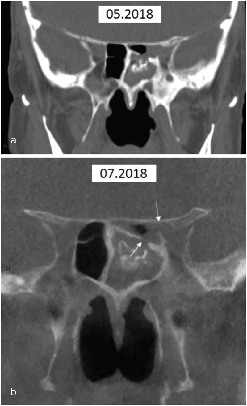
Figure 9.

CT of case 3
(a) Coronal multi-slice CT. The initial examination demonstrates bone thickening of the left ethmoid sinus and mucosal thickening embedding mineralised sinus contents. Disruption of bone is not seen; (b) Coronal cone beam CT. This confirms the findings seen in 9A but also demonstrates disruption of bone communicating with the optic canal (arrows), which were retrospectively present in 9A.
