
Figure 11.
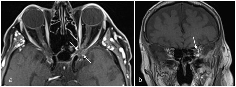
MRI of case 3 after 1 year of follow-up
(a) Gadolinium-enhanced axial T1-weighted image with fat suppression showing the absence of aspergilloma or sinusitis. Secondary optic nerve atrophy is present (arrows); (b) Gadolinium-enhanced coronal T1-weighted image with fat suppression showing the absence of inflammation and secondary optic nerve atrophy (arrow).