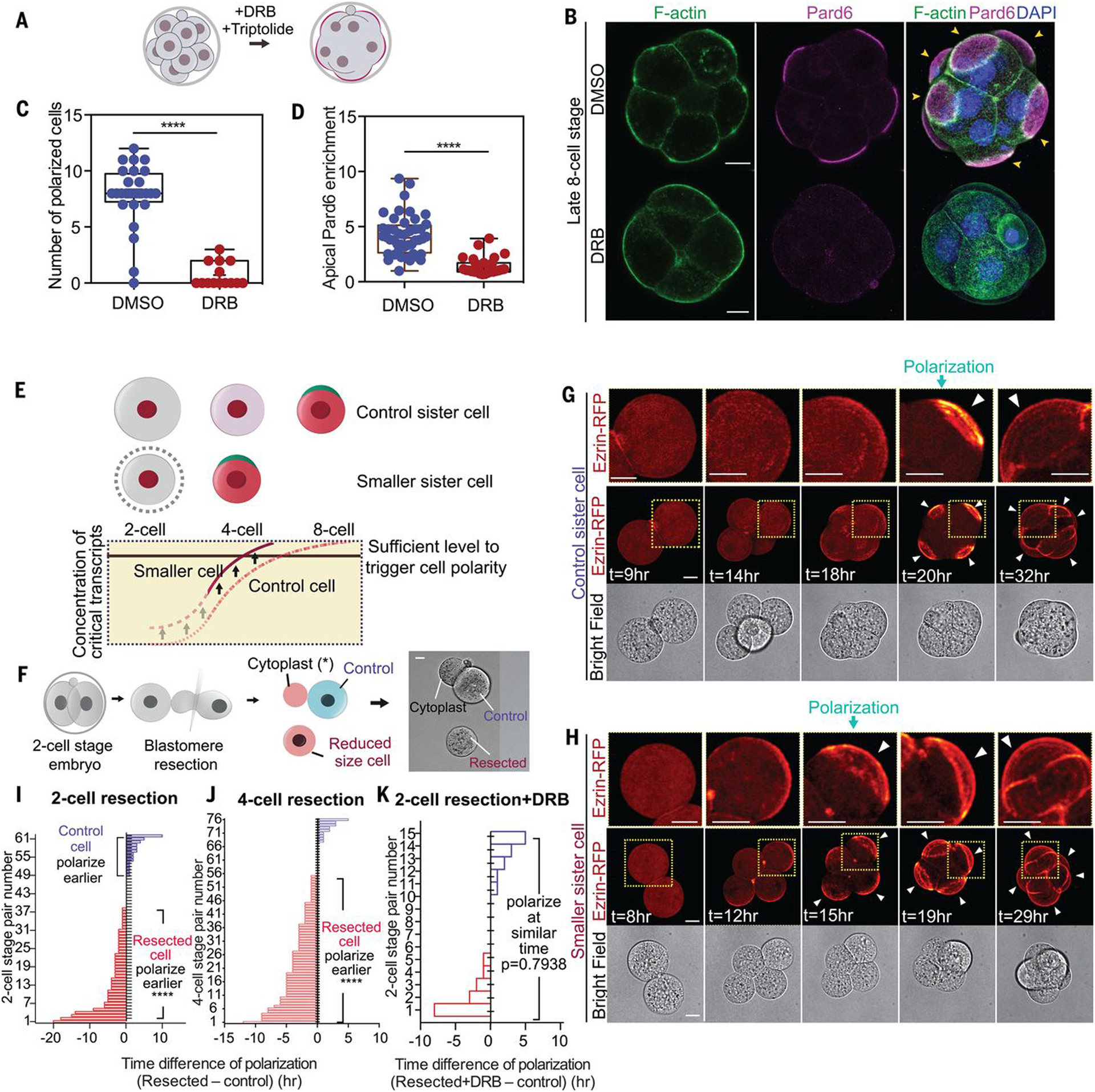
Fig. 1. The dependency of polarization on nascent transcripts.

(A) Scheme indicating inhibitor treatments. (B) Dimethyl sulfoxide (DMSO)–treated (control) or DRB-treated 8- to 16-cell embryos were analyzed for the localization of F-actin, Pard6, and DNA. Arrowheads indicate the apical domain. (C) Polarized cell number in DMSO (control) or DRB-treated embryos. ****P < 0.0001, Mann-Whitney U test. N = 2 experiments. (D) Apical enrichment of Pard6 (see methods) in 8- to 16-cell-stage cells treated with DMSO (control) or DRB. ****P < 0.0001. Mann-Whitney U test. (E) Scheme of the hypothesis: Newly synthesized factors important for polarization accumulate up to a point at which polarization is induced at the eight-cell stage. Decreasing the cell size elevates the concentration of such factors, leading to an advance in polarization timing. (F) Scheme showing the blastomere resection procedure. (G and H) Time-lapse of control or smaller sister blastomeres from the experiment described in (F). Arrowheads indicate the apical domain. Dotted yellow squares indicate the magnified regions (top row). (I and J) Polarization time difference (see methods) between smaller and control sister blastomeres from (F) or fig. S2F; each bar represents one comparison. Smaller cells polarize earlier in the significant majority of cases. N = 13 experiments for (I), N = 6 experiments for (J). ****P < 0.0001, one-sample t test, hypothetical mean = 0. (K) Polarization time difference between control and smaller DRB-treated sister cells, from experiments in fig. S2L. Each bar represents one comparison. Pulsed transcription inhibition prevents the early polarization of smaller cells. N = 3 experiments. One-sample t test, hypothetical mean = 0. Arrowheads indicate the apical domain. Scale bars, 15 μm.
