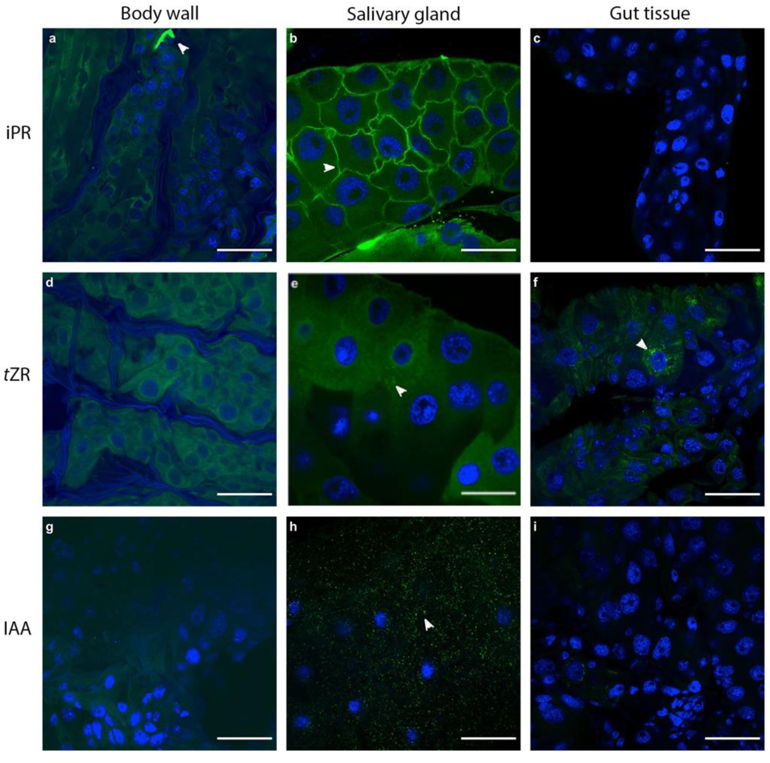
Fig 1.

Localization of cytokinin and auxin antibodies, and co-localization of Wolbachia sp. with DAPI nuclear stain. Immunolocalization using rabbit anti-iPR, tZR and IAA antibodies in E. solidaginis tissues with FITC-conjugated secondary polyclonal goat anti-rabbit antibodies (green). Localization of iPR in a) body wall, b) salivary gland tissue, and c) gut tissue. Localization of tZR in d) body wall, e) salivary gland tissue, and f) gut tissue. Localization of IAA in g) body wall, h) salivary gland tissue, and i) gut tissue. DAPI stain for cell nuclei and for all nucleic acids (blue) showed a lack of bacteria in these tissues based on the size of stained particles. DAPI fluorescence has been enhanced with brightness and contrast using ImageJ to identify all DAPI stain. All images were taken under 40x oil magnification. Scale bar for all images is 50 μm.
