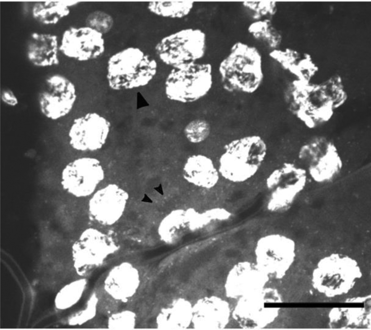
Fig 2.

DAPI stained E. solidaginis ovaries show cell nuclei (large arrowhead) and smaller flecks consistent with Wolbachia sp. chromosomes (small arrowheads). Image taken with a 40x oil-immersion lens. DAPI fluorescence has been enhanced with brightness and contrast in ImageJ to emphasize presence of small fleck staining. Scale bar for image is 50 μm.
