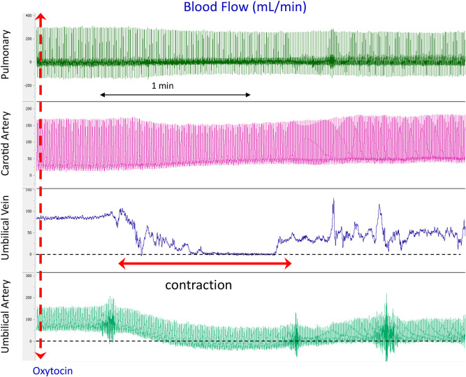
Fig 1. Physiological recordings of blood flows in the left pulmonary artery (PA), carotid artery, umbilical vein (UV) and umbilical artery (UA) before and immediately after oxytocin administration (iv) to the ewe in a newborn lamb prior to UCC and before ventilation onset.
Note that during the contraction, UV flow decreases to 0, UA flow decreases and reverses during diastole (indicated by diastolic flows below zero) and CA flow increases.

