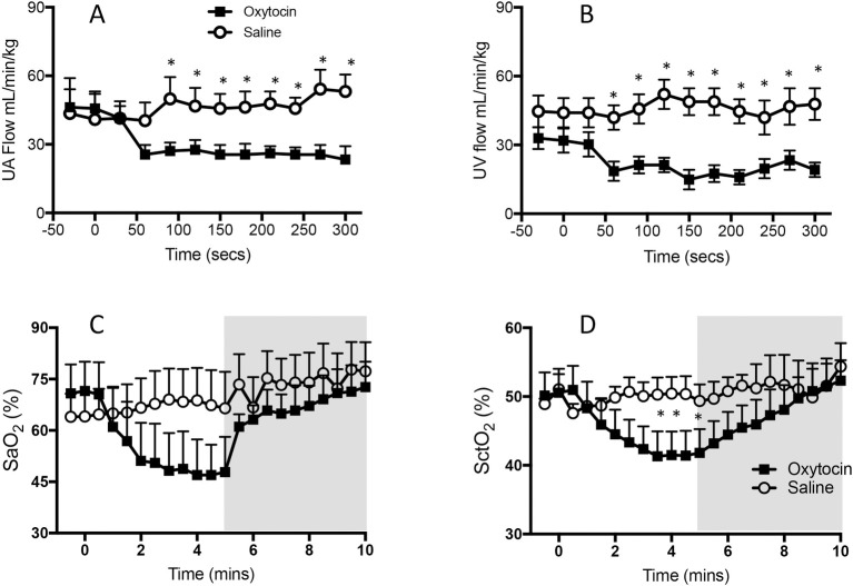
Fig 2.
Changes in (A) mean (± SEM) umbilical arterial (UA) blood flow, (B) venous (UV) blood flow (C) arterial oxygen saturation (SaO2) and (D) cerebral tissue oxygenation (SctO2) levels in RU486 plus saline treated lambs (open cycles) and RU486 plus oxytocin treated lambs (closed squares). Samples were collected during the first 5 minutes after oxytocin/saline administration and before ventilation onset (A & B) and for 5 minutes after ventilation onset (indicated by shaded area; C & D). Data from saline-infused control animals (no RU486) have been omitted for clarity.

