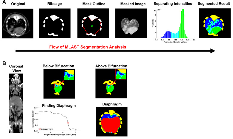
Fig 2. Schematic of MLAST analysis.
(A) Depiction of MLAST’s segmentation algorithm. The original microCT 2D image contains the densities of various tissues observed in a thoracic scan. The ribcage provides a high contrast for thresholding. The interior points of the rib regions are used to create a mask of the thoracic cavity. The densities of the voxels within the masked image are segmented with a k-means clustering algorithm and the intensities are classified into 3 tissue types: soft tissue (green), lung (blue), and intermediate (cyan). The final segmented result is shown in 2D and applied to all slices in the z-stack. (B) Depiction of how MLAST detects the cranial and caudal boundaries of the thoracic cavity. The cranial boundary is determined by the bifurcation of the trachea. Below the bifurcation, the two tracheal regions (colored blue and shown in inset) are separate. Tissues above the bifurcation, where the two regions are no longer separable, are excluded from the final segmentation. On the caudal end, the diaphragm is segmented based on density changes in the z-trace of each voxel. The resulting diaphragm (red) is removed from the thoracic counts of soft tissue, lung, and intermediate.

