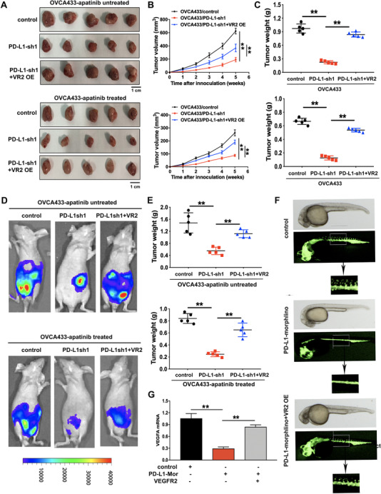
FIGURE 6.

The effects of PD‐L1 knockdown and VEGFR2 overexpression on the progression and angiogenesis of ovarian cancer in vivo. (A) Representative images of subcutaneous xenograft tumors formed by OVCA433/control, OVCA433/PD‐L1‐sh1, and OVCA433/PD‐L1‐sh1 + VEGFR2 overexpression (VR2 OE) cells in nude mice with or without apatinib treatment. (B) Growth curve of the xenograft tumors in mice with or without apatinib treatment. (C) The average tumor weight of nude mice with or without apatinib treatment. (D) Representative images of intraperitoneal xenograft tumors in nude mice with or without apatinib treatment. (E) The average weight of intraperitoneal xenograft tumors in nude mice. (F) Zebrafish model. Zebrafish embryos were injected with PD‐L1 morphlino or VEGFR2 cDNA recombinant plasmid. (G) The mRNA expression levels of VEGFA assessed by qRT‐PCR assay in zebrafish model. **P < 0.01. Abbreviations: PD‐L1: programmed cell death‐ligand 1; PD‐L1‐sh1/2: PD‐L1 short‐hairpin RNA 1/2; VR2‐OE: vascular endothelial growth factor receptor‐2 overexpression; PD‐L1‐Mor: PD‐L1 morphlino; qRT‐PCR: quantitative real‐time polymerase chain reaction
