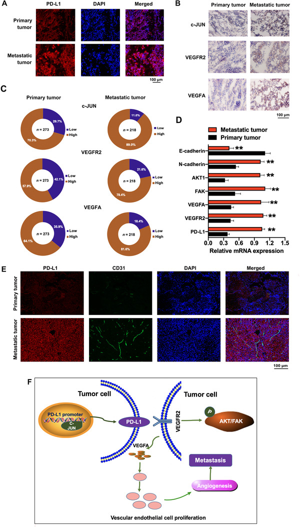
FIGURE 7.

The PD‐L1, c‐JUN, VEGFR2, and VEGFA expression in primary and metastatic ovarian cancer tissues. (A) Immunofluorescence staining of PD‐L1 in primary and metastatic ovarian cancer tissues. (B) Immunohistochemical staining of c‐JUN, VEGFR2, and VEGFA in primary and metastatic ovarian cancer tissues. (C) The statistical results of the immunohistochemical staining of c‐JUN, VEGFR2, and VEGFA. (D) The mRNA expression levels of genes associated with cell migration and angiogenesis were tested by qRT‐PCR assay. (E) Immunofluorescence staining of PD‐L1 and vascular endothelial cell‐specific marker CD31 in primary and metastatic ovarian cancer tissues. (F) Schematic model showing the role of the c‐JUN/PD‐L1/VEGFR2 signaling axis in the regulation of angiogenesis. Abbreviations: PD‐L1: programmed cell death‐ligand 1; DAPI: 4′,6‐diamidino‐2‐phenylindole; qRT‐PCR: quantitative real‐time polymerase chain reaction; AKT1: protein kinase B; FAK: focal adhesion kinase; VEGFA: vascular endothelial growth factor A; VEGFR2: vascular endothelial growth factor receptor‐2
