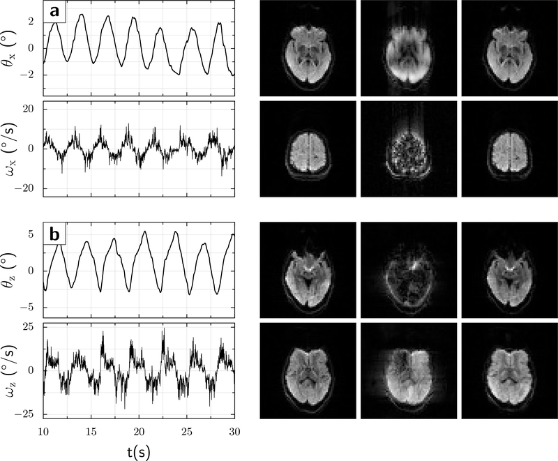
Figure 5:
A volunteer moved his head quasi-periodically in two separate experiments. (a) nodding in the up-down direction (x-rotation), turning points θx ≈ ±3° (solid red line), and peak rotational velocity ≈ 7.2°/s (solid blue line). (b) Left-right head rotation (z-direction), turning points θz ≈ ±5.5° (solid red line), and peak rotational velocity ≈ 13°/s (solid blue line). The images show 2 representative slices for each motion condition, as follows: Left column: No movement; Center column: Movement but motion correction Off; and Right column: Movement with motion correction On. In both instances, the scans with motion but no intra-sequence updates show markedly more motion artifacts or signal losses.

