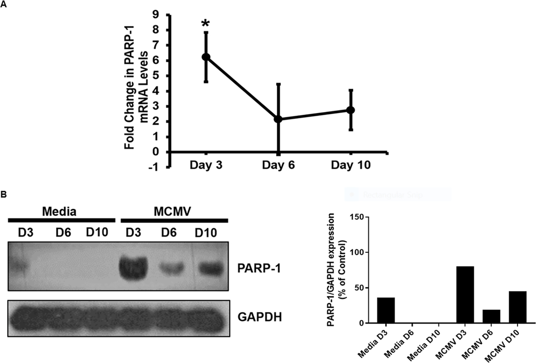
Figure 1.
Detection of PARP-1 mRNA (Fig 1A) and protein (Fig 1B) within MCMV-infected eyes of MAIDS mice at 3, 6, and 10 days after subretinal MCMV inoculation compared with mock-infected control eyes. PARP-1 mRNA expression values as determined by qRT-PCR assay represent means from n = 3 – 5 mice per group from two independent experiments, and error bars indicate standard errors of the mean (SEM). Statistical significance between groups per day (*, p < 0.05) was determined by Student’s t test. PARP-1 protein detection represents pooled whole eyes (n = 3 – 5 mice per group).

