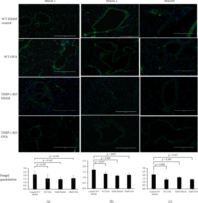
Figure 3.

Membrane-bound mucins MUC1, MUC 2, and MUC4 expression in lung tissue obtained from OVA-sensitized TIMPKO, OVA-sensitized TIMPKO, OVA-sensitized WT, and WT-SHAM mice. Representative images of immunofluorescent staining of MUC1 (a), MUC2 (b), and MUC4 (c). The fluorescent intensity was quantitively assessed via the ImageJ software, with the representative histogram describing the data obtained from n = 3 separate experiments with WT-SHAM control.
