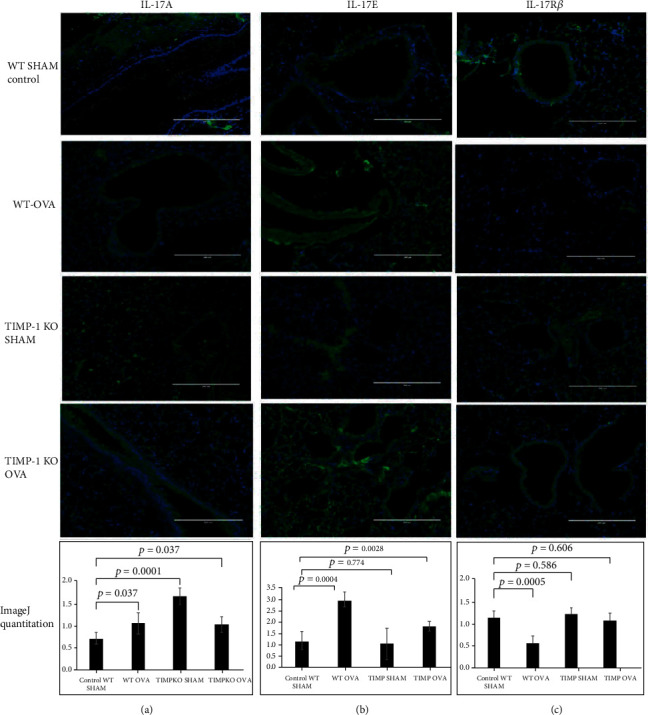
Figure 5.

IL-17A, IL-17Rβ, and IL-17E expression in lung tissue obtained OVA-sensitized TIMP-1 K/O, OVA-sensitized TIMPKO, OVA-sensitized WT, and WT-SHAM mice. Representative images of immunofluorescent staining of IL-17A (a), IL-17E (b), and IL-17Rβ (c). The fluorescent intensity is quantitated using the ImageJ software, and the representative histogram derived from n = 3 separate experiments with WT-SHAM as control.
