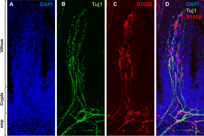
Figure 2.
Mucosal enteric glial cell (mEGC) network is present in the human fetal gut. Cryosections of human fetal small intestine were stained with DAPI (A), anti Tuj1 (B) and anti S100β (C) antibodies. Individual channels are displayed in (A–C) and merged in (D). Innervation (B) and mEGC network (C) of the human fetal gut mucosa (villous and crypts) and submucosal plexus (SMP) are demonstrated at 16 weeks gestational age. Confocal images of fetal gut cryosections were acquired with Leica TCS SP5 with a DN6000 microscope assisted by the LAS AF software. All images were processed with ImageJ (Wayne Rasband, NIH) using 3-D reconstructions and opacity mode. Original magnification X40.

