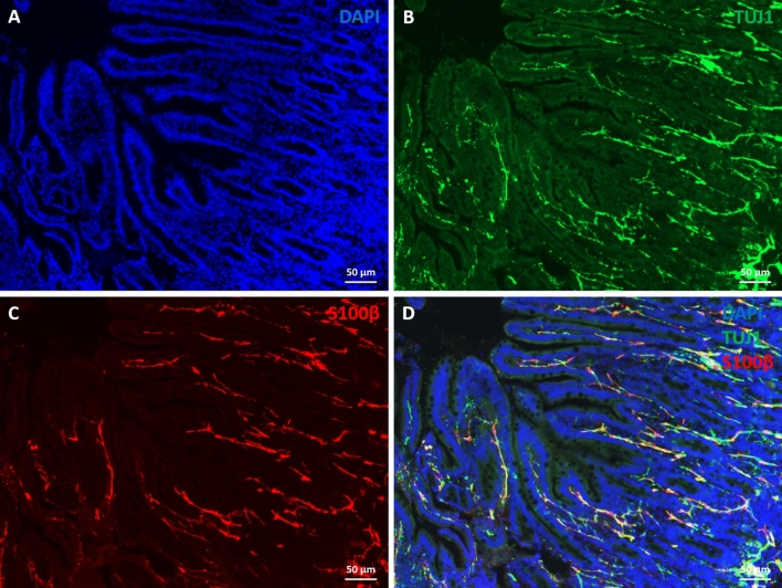
Figure 3.
Presence of mucosal enteric glial cells in human gut xenograft, despite the absence of luminal microbiota. Cryosections of human fully developed gut xenografts were stained with DAPI (A), anti Tuj1 (B) and anti S100β (C) antibodies. Individual channels are displayed in A–C and merged in D. Confocal images of gut cryosections were acquired with Leica TCS SP5 with a DN6000 microscope assisted by the LAS AF software. All images were processed with ImageJ (Wayne Rasband, NIH) using 3-D reconstructions and opacity mode. Scale bars 50 µm.

