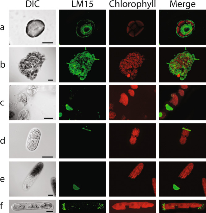
Fig. 4. XyG Immunolabeling localizes to the cell wall of several CGA species from the three later evolved classes of CGA.

a Coleochaete orbicularis XyG LM15 labeling is strong in the remnants of the zoospore cell wall and in the periphery of the newly formed thallus. b The peripheral cells of thalli as well as elongating hairs also show distinct labeling. c Coleochaete nitellarum XyG LM15 labeling is strong at the tips of growing filament structures. In elongating d Cylindrocystis brebissonii, e Netrium digitus and f Mesotaenium caldariorum cells, XyG specific labeling with LM15 is restricted to zones in the cell wall near one end or both ends of the cell, likely involved in cell expansion. DIC: light image, LM15: TRITC (green), chlorophyll autofluorescence (red). Merge: LM15 and chlorophyll autofluorescence. Scalebars as indicated in the DIC images: a 10 µm and b–f 20 µm.
