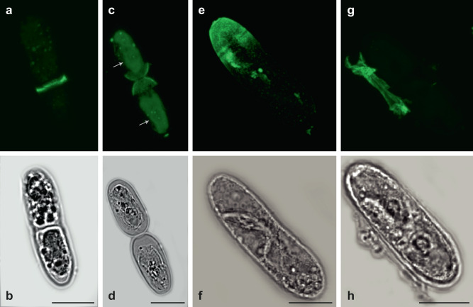
Fig. 5. XyG production in Mesotaenium caldariorum during semicell morphogenesis.
Immunolabelling of cell cycle synchronized Mesotaenium caldariorum cells during semicell morphogenesis. a, b LM15 binding was found in the isthmus when cells were starting to divide. c, d LM15 is evident in the ends of daughter cells near the division zone (while chlorophyll is also evident and colored green in the middle of the two daughter cells as indicated by arrows) e, f cell expansion is almost completed of the new Mesotaenium caldariorum cells and LM15 labeling is very clear in the newly formed “primary cell wall”. g, h Cell division and expansion is completed and the “primary cell wall” is cast off as a single piece depicted here, including the LM15 labeling. Top panel: LM15 (TRITC) (green), lower panel: DIC light image. c Chlorophyll autofluorescence (green), in the middle of the cells. Scalebars as indicated in the DIC images: 20 µm.

