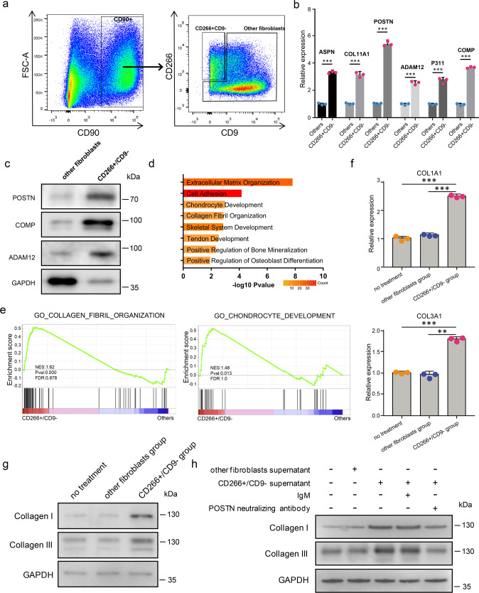
Fig. 5. The supernatant of mesenchymal fibroblasts promotes collagen expression in keloid fibroblasts.
a Isolation of CD266+CD9− and other fibroblasts from keloid dermis by flow cytometry. b qRT-PCR assay of some mesenchymal fibroblast marker genes expression in CD266+/CD9− fibroblasts and other fibroblasts from keloid dermis. Data are presented as mean values ± SD (n = 3 biologically independent experiments.). Two-sided unpaired t-test, ***P < 0.001(ASPN:P = 0.00003, COL11A1:P = 0.00019, POSTN:P = 0.00004, ADAM12:P = 0.00015, P311:P = 0.00019, COMP:P = 0.00002). c Western blot assay of mesenchymal fibroblast marker genes expression in CD266+/CD9− fibroblasts and other fibroblasts from keloid dermis. The experiments were repeated three times with three different fibroblast donors, and here a representative result was shown. d GO Biological Process enrichment analysis of differentially expressed genes between keloid CD266+/CD9− fibroblasts and other fibroblasts. e GSEA enrichment plots for representative signaling pathways upregulated in keloid CD266+/CD9− fibroblasts compared to other fibroblasts. (NES normalized enrichment score, corrected for multiple comparisons using FDR method, P-value were showed in plots). f, g Keloid other fibroblasts were treated with the supernatant of CD266+/CD9− or other fibroblasts. The expression of collagen I and collagen III was analyzed by qRT-PCR (f) or western blot (g). Data are presented as mean values ± SD (n = 3 biologically independent experiments). Two-sided unpaired t-test, **P < 0.01, ***P < 0.001. (COL1A1: P = 0.000009 and P = 0.000015, respectively; COL3A1: P = 0.00006 and P = 0.00214, respectively). The experiments were repeated three times with three different fibroblast donors, and here a representative result was shown. h Keloid other fibroblasts were treated as indicated in the figure. The expression of collagen I and collagen III was analyzed by western blot. The POSTN antibody neutralizing experiments were repeated three times with three different fibroblast donors, and here a representative result was shown.

