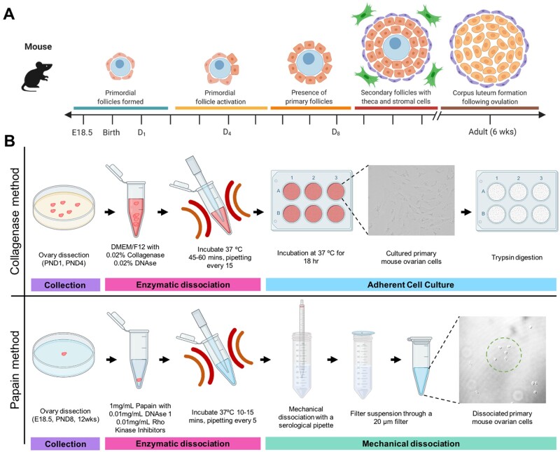
Figure 1.
Schematic diagram showing the timeline of ovarian follicle development in the mouse and the two dissociation protocols to retrieve somatic cells from these ovarian timepoints. (A) Relative timeline showing the key stages of folliculogenesis in embryonic mice (E = embryonic day) and post-natal mice (PND = days post birth). Germ cell cysts, also known as nests, breakdown to form primordial follicles (teal bar), containing an oocyte (blue) and pre-granulosa cells (orange), then the initial wave of primordial follicle activation occurs (yellow bar) which sets the ovarian reserve, leaving a proportion of primary follicles (orange bar) present in the ovary. Primary follicles then continue through folliculogenesis to form secondary follicles (red bar), which are surrounded by theca cells (purple) and stromal cells (green). Once the mice become sexually mature, around 6 weeks of age, corpus luteum structures form following ovulation, and the granulosa cells become luteinised and produce hormones (brown bar). Figure adapted from (Sarraj and Drummond, 2012), figure made using Biorender.com. (B) Schematic diagrams of the key steps in the collagenase and papain methods. Both methods involve collection steps (purple bar) and enzymatic dissociation steps (pink bar). The collagenase method uses differential cell adhesion in culture to select for granulosa cells (teal bar). In contract, the papain method using mechanical dissociation and filtration to enrich for somatic cells (blue bar). Figure made using Biorender.com.

