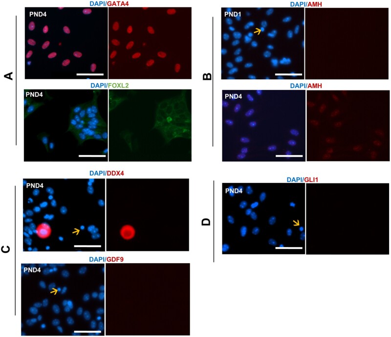
Figure 3.
Fluorescence images for collagenase dissociation method. (A) Immunofluorescent localisation of ovarian cell markers counterstained with blue nuclear marker (4′‐6‐diamidino‐2‐phenylindole (DAPI)) and the granulosa cell markers (GATA4 and FOXL2), (B) differentiating granulosa cell marker (AMH) in post-natal day (PND) 1 and PND4 cells dissociated via collagenase, (C) oocyte markers (DDX4 and GDF9), and (D) theca cell marker (GLI1). Arrows indicates apoptotic cells. All experiments performed in PND1 and PND4 n = 3 for each group, representative images taken at PND4 stage except where indicated. Images taken at 40× magnification, scale bar equivalent to 50 μm.

