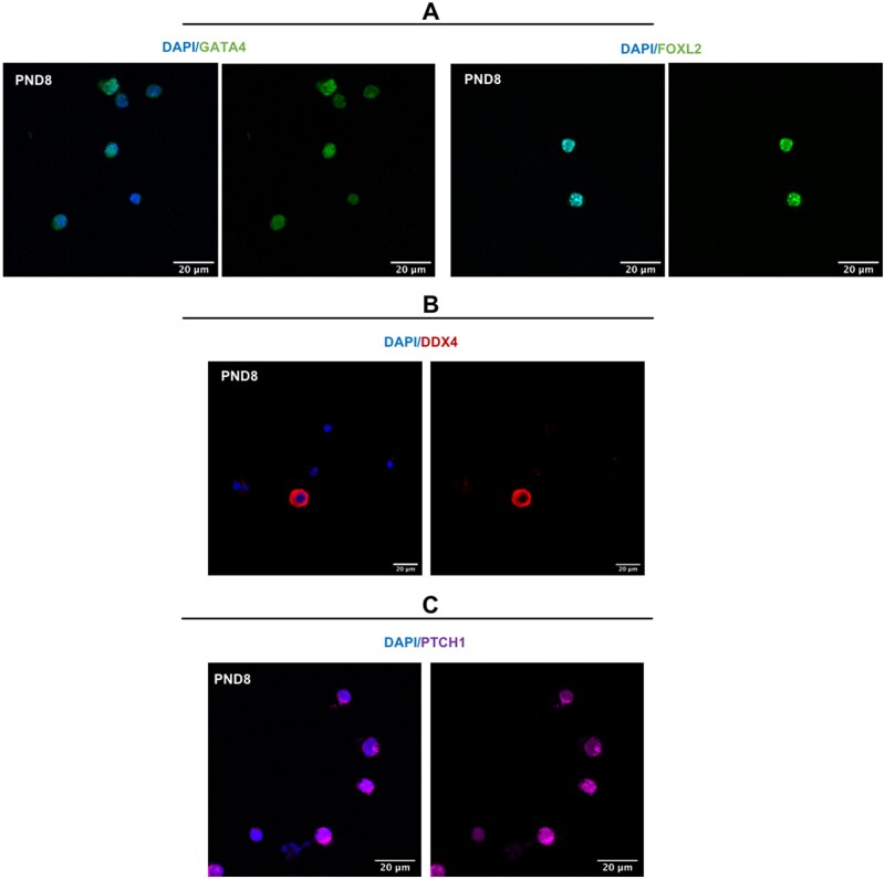
Figure 5.
Fluorescence images for papain dissociation method. Immunofluorescent localisation of ovarian cell markers counterstained with blue nuclear marker (4′‐6‐diamidino‐2‐phenylindole (DAPI)): (A) granulosa cell markers (GATA4 and FOXL2), (B) oocyte marker (DDX4), and (C) theca cell marker (PTCH1). All experiments performed on dissociated ovarian cells from PND8 ovary (n = 3), and representative images shown. Images taken at 60× magnification, scale bar 20 μm.

