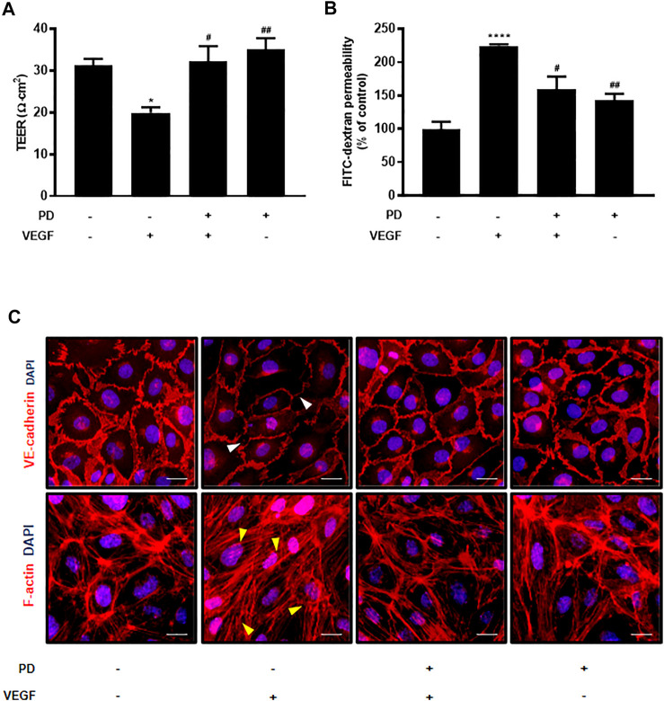
FIGURE 1.
Primaquine diphosphate (PD) blocks VEGF-induced endothelial permeability and stabilizes junctional proteins. HUVECs were starved and treated with or without PD (5 μM, 30 min) before stimulation with VEGF (30 ng/ml, 30 min). PD blocked both TEER decline (A) and increased FITC-dextran transendothelial permeability (B) induced by VEGF. TEER was measured using Millicell ERS-2 (Millipore). For the permeability assay, FITC-dextran was added to the upper chamber. The absorbance of the solution in the lower chamber was measured at 492 nm (excitation) and 520 nm (emission) using a FLUOstar Omega microplate reader. n ≥ 3 independent experiments. (C) HUVECs were starved and treated with or without PD (5 μM, 30 min) before stimulation with VEGF (50 ng/ml, 30 min). Cells were fixed, permeabilized, and immunostained for VE-cadherin and F-actin. White arrow indicates an attenuated VE-cadherin expression and yellow arrow shows stress fiber formation. All data are presented as means ± SEM, *p < 0.05, ****p < 0.0001 vs. control group; #p < 0.05, ##p < 0.005 vs. VEGF treatment group. Scale bar = 20 μM.

