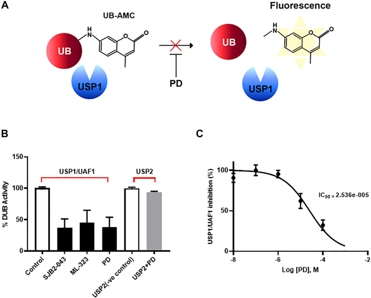
FIGURE 4.
PD specifically inhibited USP/UAF1 activity. (A) Schematic representation of Ub-AMC assay. USP1 removes ubiquitin from its substrate Ub-AMC, and fluorescent AMC is measured. (B) rUSP1/UAF1 complex or rUSP2 were incubated with control, USP1 inhibitor SJB2-043, ML-323, or PD for 30 min at 37°C, followed by assessment of DUB activity using Ub-AMC assay. (C) Progress curve for USP1/UAF1 activity on PD against Ub-AMC. The graph represents the average of three independent experiments with calculated SEM. The experiment was conducted at least four times.

