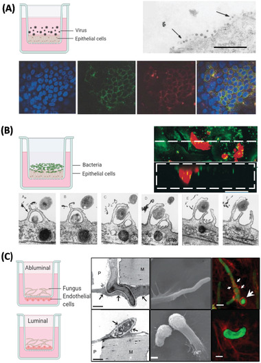
Figure 3.

Examples of in vitro models used to study entrance and virulence mechanisms of A) viral, B) bacterial, and C) fungal pathogens. A) Apical entry and release of severe acute respiratory syndrome‐associated coronavirus in polarized Calu‐3 lung epithelial cells. Above: Transmission electron microscopy of release of SARS‐CoV virons from the apical surface of polarized Calu‐3 cells. Below: Colocalization of ACE‐2 and viral antigen in infected Calu‐3 cells, both ACE‐2 (green) and viral antigen (red) could be detected in infected cells. Importantly, both ACE‐2 and viral antigen appeared to colocalize in infected cells (yellowish). Reproduced with permission.[ 92 ] Copyright 2005, ASM. B) Infection of primary human bronchial epithelial cells by Hemophilus influenzae. Above: Images collected by dual‐wavelength CLSM of cells infected for 3 h; colocalization of airway nuclei, bacteria (green) and vacuoles (red) can be seen in yellow, suggesting bacteria have been taken into the cells. Scale bar is 50 µm. Below: The series (A through F) demonstrates lamellipodia surrounding bacteria (black arrow) at the surface of a submerged airway cell culture after 4 h of infection. Reproduced with permission.[ 141 ] Copyright 1999, ASM. C) Polarized response of endothelial cells to invasion by Aspergillus fumigatus. A. fumigatus hyphae invade the abluminal and luminal surface of endothelial cells by different mechanisms. Above: Hyphae invading the abluminal surface of endothelial cells, Arrows indicate an endothelial cell that is being invaded by a hypha. Below: Hyphae invading the luminal surface, arrows indicate endothelial cell pseudopods. Hyphae are shown in green and microfilaments in red. Bars represent 5 µm. Reproduced with permission,[ 142 ] Copyright 2009, Wiley. The inset cartoon schematics represent the type of model chosen. Cartoon insets created using BioRender.com.
