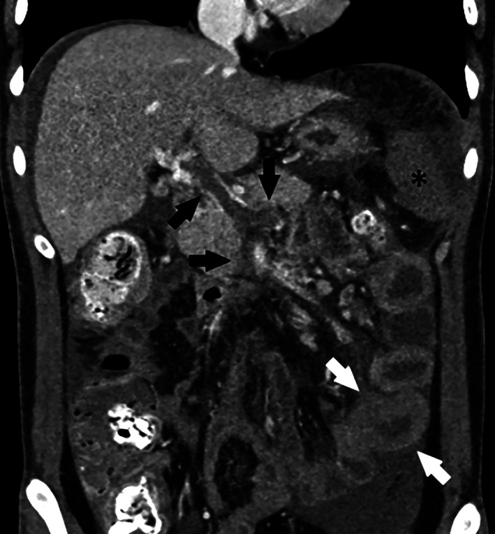
FIGURE 1.

Pre‐TIPS coronal image from CT performed on Day 4 of hospitalization showing portal, SMV and splenic vein thrombosis (black arrows), bowel wall thickening (white arrows), and splenic hypoenhancement (asterisk) worse compared to the CT on admission
