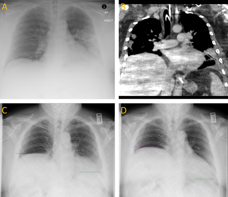
Figure 1.
(A) Initial anteroposterior (AP) chest radiograph demonstrating left mid-zone and lower zone consolidation, with both haemidiaphragms in a conventional position. (B) Coronal CT thorax showing bilateral multifocal peripheral consolation with a raised right haemidiaphragm and no mediastinal mass. (C, D) Still frames from a posteroanterior (PA) DCR during sniff test at expiration (C) and inspiration (D) showing further elevation and paradoxical motion of the right haemidiaphragm. Resolution of the lung parenchymal changes has occurred. DCR, dynamic chest radiography.

