Figure 4.
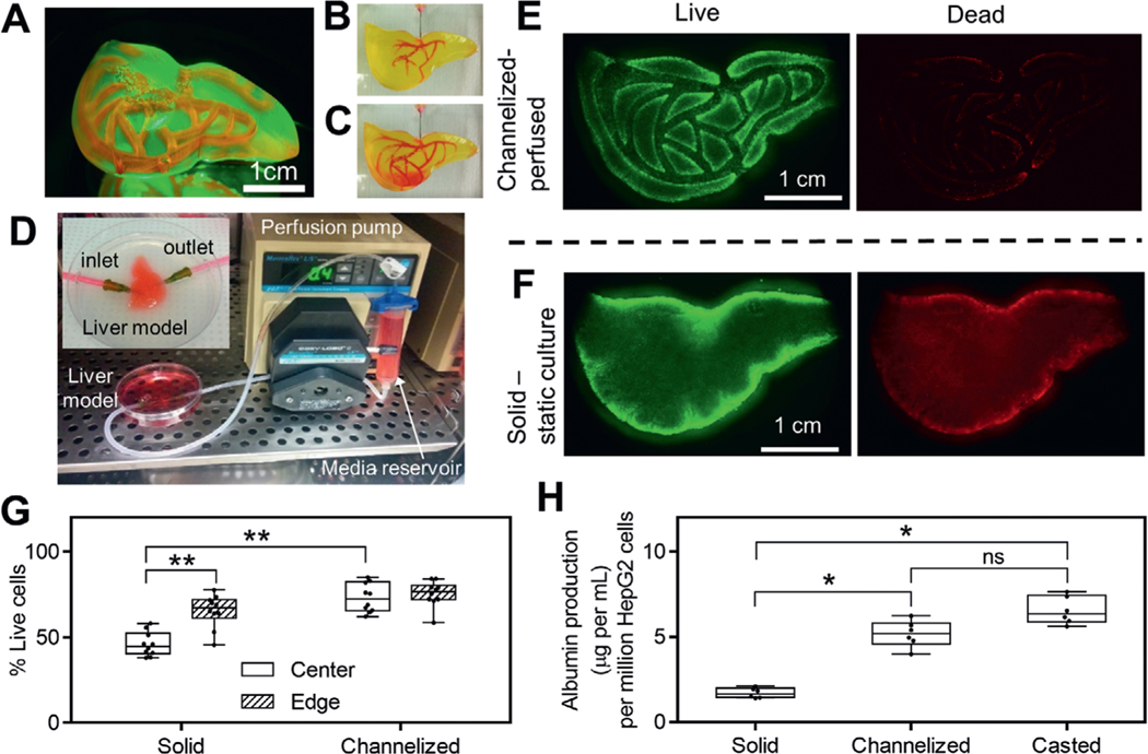
Media perfusion in printed channels helps to maintain long-term cell functions. A) A liver model with smooth surface and monolithic, translucent hydrogel body was printed using 15% PEGDA 4 kDa. Vascular channel network was filled with rhodamine B and visualized under fluorescence. At the beginning B) and at the end C) of rhodamine B injection into prefabricated, vascular-like channels. D) Experimental setup of the perfusion system in an incubator. Inset shows enlarged view of the perfusion chamber for liver model. Inlet and outlet of the channel network are connected to perfusion tubing through 18 gauge needles. Fluorescent images of live and dead cells in channelized, perfused liver model (E) and solid, F) statically cultured liver model after 3 d of culture. G) Measured cell viability in channelized and solid liver models at both the center and edge regions, n = 10. H) Albumin production for channelized liver model, solid liver model and casted model after 6 d of culture, n = 6. Cell-laden liver models in E–G) were printed using 15% PEGDA 4 kDa conjugated with 10 × 10−3m RGD and liver model in H was printed using 8% GelMA plus 5% PEGDA 8 kDa. All box plots with whiskers represent the data distribution based on five number summary (maximum, third quartile, median, first quartile, minimum). **, p < 0.001; *, p < 0.05; determined by nonparametric unpaired t-test.

