Figure 4.
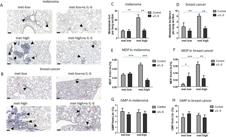
IL-6 pathway blockade inhibits metastasis. Mice were implanted with met-low or met-high melanoma or breast cancer cells. One week later, mice were treated with IgG (control) or anti-IL-6 antibodies twice weekly. At end point, mice were sacrificed, lungs were removed, and bone marrow was harvested (n=5–8 mice/group). (A–B) Representative images of lung sections from melanoma (A) and breast cancer (B) are shown, bar=100 µm. Arrows indicate metastatic foci. (C–D) Metastatic foci per lung section were quantified (n=5–7 sections/mouse) for melanoma (C) and breast cancer (D). (E–F) MDP levels in BM of melanoma (E) and breast cancer (F) were assessed by flow cytometry. (G–H) GMP levels in BM of melanoma (G) and breast cancer (H) were assessed by flow cytometry. Statistical significance was assessed by one-way analysis of variance, followed by Tukey post hoc test when comparing more than two groups or unpaired two-tailed t-test when comparing two groups. Asterisks represent significance from control, unless indicated otherwise in the figure. Significant p values are shown as *p<0.05; **p<0.01; ***p<0.001. BM, bone marrow; GMP, granulocyte-monocyte progenitor; IL-6, interleukin 6; MDP, monocyte-dendritic progenitor; met-high, high metastatic potential; met-low, low metastatic potential.

