Figure 6.
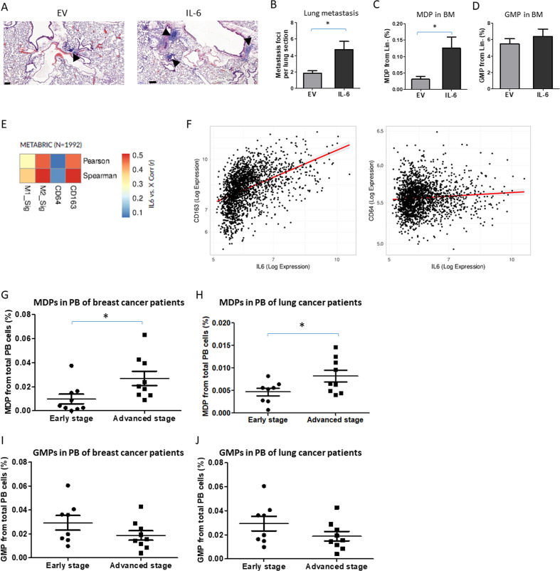
Increased immunosuppressive macrophages and MDPs correlate with IL-6 expression and aggressive tumors. (A–D) C57BL/6 mice aged 8–10 weeks were implanted with B16 IL-6 overexpressing cells or with corresponding control EV cells. At end point (day 18), mice were sacrificed, lungs were removed, and the bone marrow was harvested (n=4–6 mice/group). (A) Representative images of lung sections are shown, bar=100 µm. Arrows indicate metastatic foci. Metastatic foci per lung section were quantified (n=4–6 sections/mouse). (C) MDP and (D) GMP levels in BM were assessed by flow cytometry. (E) Heat-map of linear correlations in R values between IL-6 expression and M1 or M2 gene signatures as well as single genes CD64 or CD163 representing M1 and M2 subsets, respectively. The data were obtained from human breast cancer samples using the METABRIC data set. (F) Scatter graphs of IL-6 expression in correlation with CD64 or CD163 in human breast cancer samples (METABRIC data set). (G–J) MDP (G, H) and GMP (I, J) levels were analyzed in peripheral blood of patients with breast (G, I) and lung (H, J) cancer segregated based on early and advanced stage disease. Statistical significance was assessed by unpaired two-tailed t-test. Significant p values are shown as *p<0.05. BM, bone marrow; EV, empty vector; GMP, granulocyte-monocyte progenitor; IL-6, interleukin 6; MDP, monocyte-dendritic progenitor; PB, peripheral blood.

