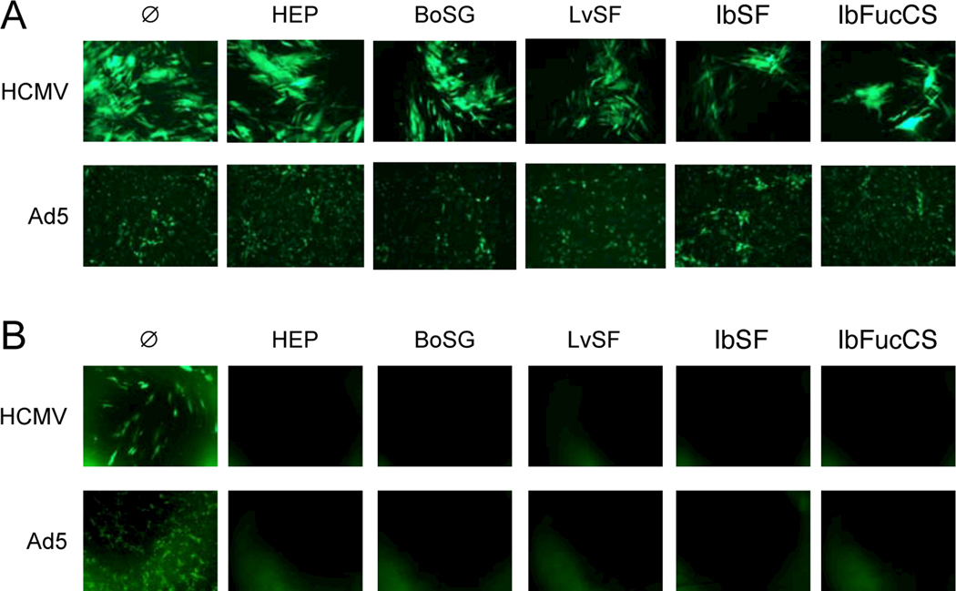
Fig. 5. Treatment/removal studies.
(A) Confluent monolayers of MRC-5 fibroblasts (top) or ARPE-19 epithelial cells (bottom) in 96-well plates were treated with medium (Ø) or 150 μg/mL heparin (HEP), BoSG, LvSF, IbSF, or IbFucCS for one h then cells were washed three times with medium and infected with GFP-tagged HCMV BADr (100 PFU/well) or GFP-tagged Ad5 (100 PFU/well). (B) HCMV BADr or adenovirus virions were incubated with sulfated glycans as in (A) for 1 h, then diluted 10,000-fold with culture medium to a non-inhibitory concentration (15 ng/mL). Virions with sulfated glycans were then added to MRC-5 fibroblasts (top) or ARPE-19 epithelial cells (bottom) in 96-well plates. Representative fluorescent micrographs were taken six days post infection.

