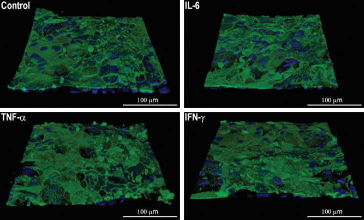
Figure 5.
Representative confocal Z-stack images showing glycocalyx staining in stratified cultures of human conjunctival epithelial cells exposed to IL-6, TNF-α, and IFN-γ (250 pg/mL) for 24 hours. Nuclei are stained blue with DAPI and glycocalyx is stained green with Alexa-488 conjugated wheat germ agglutinin lectin. Scale bar = 100 µm.

