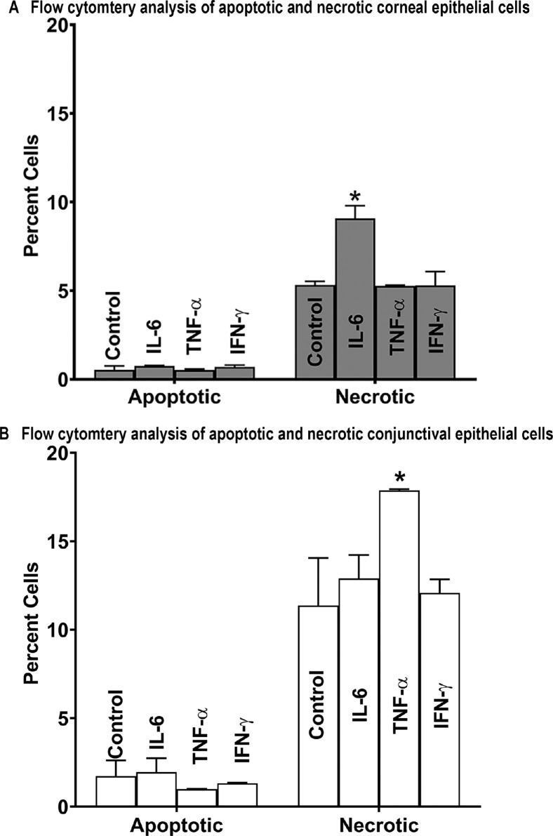
Figure 6.
Flow cytometry quantification of apoptosis and necrosis in stratified human corneal (A) and conjunctival (B) epithelial cells treatment with IL-6, TNF-α, and IFN-γ (250 pg/mL) for 24 hours. The cells were stained with annexin V/propidium iodide dual staining for flow cytometry quantification. *P < 0.05 as compared to control cells that were not exposed to any cytokine.

