
Figure 7.
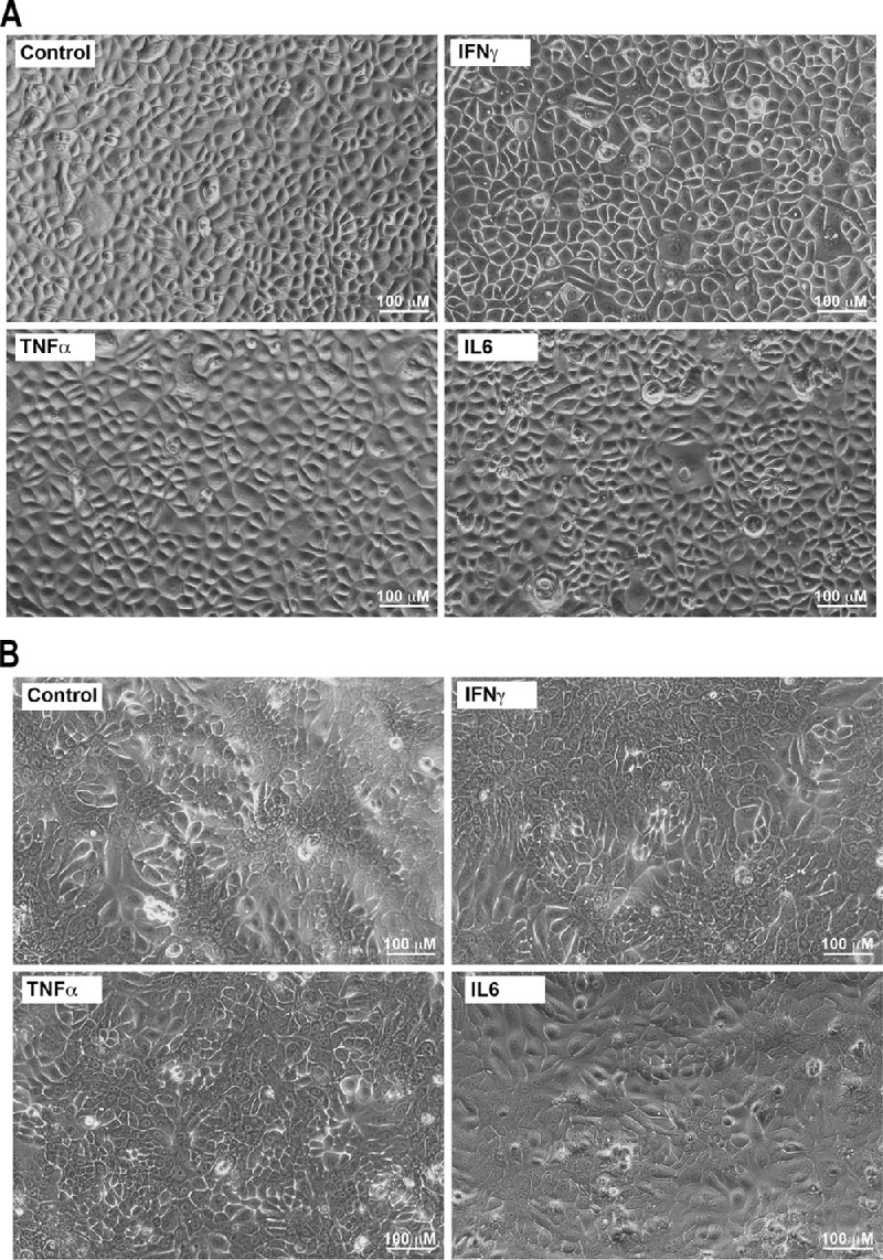
Representative phase contrast microscopy images of stratified human corneal (A) and conjunctival (B) epithelial cells exposed to IL-6, TNF-α, and IFN-γ (250 pg/mL) for 24 hours. Scale bar = 100 µm.
Official websites use .gov
A
.gov website belongs to an official
government organization in the United States.
Secure .gov websites use HTTPS
A lock (
) or https:// means you've safely
connected to the .gov website. Share sensitive
information only on official, secure websites.
Representative phase contrast microscopy images of stratified human corneal (A) and conjunctival (B) epithelial cells exposed to IL-6, TNF-α, and IFN-γ (250 pg/mL) for 24 hours. Scale bar = 100 µm.